It’s natural to feel concerned when a mole changes in size, colour, or shape, or when a new lesion appears. A professional mole check helps identify harmless moles, detect early signs of melanoma, and decide whether any lesions need monitoring or removal. At our London and Harley Street clinics, consultant dermatologists use detailed visual examination and dermoscopy to assess each mole accurately and explain whether further investigation is needed. Your appointment is calm, thorough, and focused on giving you clear answers and confidence in your skin health.
A mole should be assessed by a dermatologist if it changes in appearance, behaves differently, or simply looks unusual compared with your other moles. Most moles are harmless, but early review is essential because melanoma can develop from new or existing moles and often begins with subtle visual changes.
A mole that changes in size, colour, shape, or texture over several weeks should be assessed. Gradual evolution is one of the most reliable indicators that something needs medical review.
Any new mole appearing after the age of 30 warrants a check, especially if it looks different from your other moles.
Bleeding, itching, crusting, or becoming tender are signs the mole may be irritated or require further evaluation.
Moles that look noticeably uneven or have edges that are jagged or blurred are more likely to need medical assessment.
If you have a history of melanoma, atypical moles, or strong sun damage, regular skin checks are recommended even if a mole appears unchanged.
Melanoma symptoms can vary widely, but the key concern is any mole or pigmented mark that begins to behave differently from the rest of your skin. Melanoma often develops from an existing mole, but it can also appear as a new mark, particularly on sun-exposed areas. Changes in colour, shape, size, or surface texture are the most common warning signs, and these changes can occur gradually or over a relatively short period.
A mole that becomes darker, develops multiple colours, or shows irregular shading should be assessed promptly. Edges that begin to blur, form notches, or lose symmetry can also indicate abnormal cell activity. Some melanomas grow outwards in an asymmetric or raised pattern, while others spread deeper into the skin before becoming visible on the surface.
Symptoms aren’t always visual. Persistent itching, a new sensation of discomfort, or tenderness around a mole can signal underlying inflammation. Bleeding, crusting, or a mole that begins to ooze are more advanced warning signs and should be evaluated without delay. A mole that appears after the age of 30 and grows quickly is also considered higher risk.
Changes in sensation or texture, such as a mole becoming firm, rough, or developing a new raised area, can indicate progression beneath the skin. Melanoma can sometimes resemble a small wound that doesn’t heal normally, particularly on the legs or back.
These symptoms do not confirm melanoma, but they are medically significant and warrant assessment by a dermatologist. Early detection remains the most important factor in improving outcomes, and any noticeable or persistent change should be checked as soon as possible.
| Step | What to expect |
|---|---|
| 1. Booking and pre-assessment | You’ll book a mole or full skin check with one of our skin cancer specialists. We’ll ask brief questions about your skin type, family history of melanoma, previous sun exposure, and any moles you’re worried about. |
| 2. Specialist consultation | Your appointment is carried out by a consultant dermatologist or plastic surgeon with specific training in skin cancer. They’ll take a focused history, examine your skin, and identify any moles or lesions that need closer assessment. |
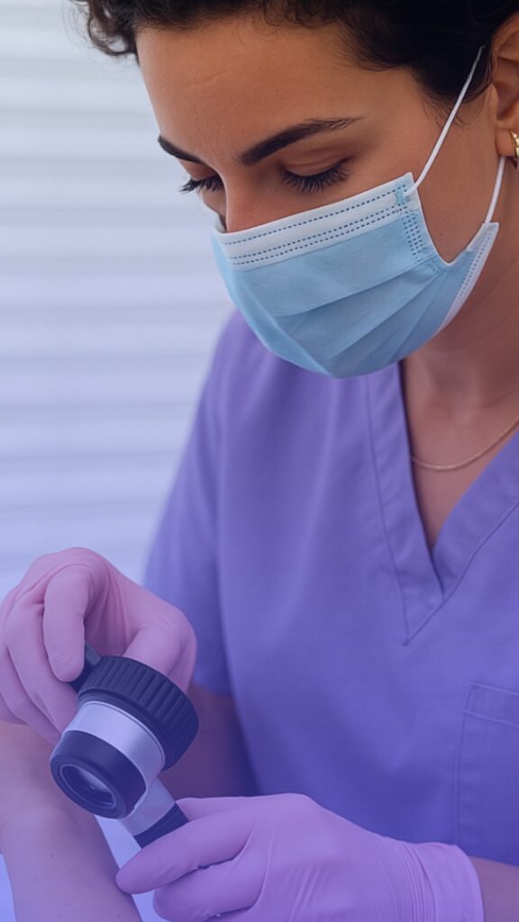
| 3. Dermoscopic assessment | Suspicious or changing moles are examined with a dermatoscope, a magnifying device that shows pigment patterns and structures not visible to the naked eye. This helps distinguish benign moles from lesions that may require biopsy or removal. |
| 4. Clinical decision | Based on the clinical and dermoscopic findings, the doctor will explain whether a mole appears benign, needs monitoring, or should be removed for safety. You’ll have the chance to ask questions about risk, next steps, and treatment options. |
| 5. Mole removal or biopsy (if needed) | When removal is recommended, the procedure is performed by a consultant plastic surgeon at our clinic. Under local anaesthetic, the mole is removed using the most appropriate technique (usually excision or shave removal), and the sample is sent for histology to rule out melanoma or other skin cancers. |
| 6. Histology and results | All medically indicated removals are analysed by a specialist pathology lab. Your results are reviewed by your consultant, who will explain the report, confirm whether further treatment is required, and arrange any follow-up if needed. |
| 7. Follow-up and ongoing monitoring | You’ll be given clear advice on self-examination, sun protection, and when to return for future checks. If you’re at higher risk of skin cancer, your doctor may recommend regular screening intervals. |
| Service | What It Involves |
|---|---|
| Full Body Mole Check | A head-to-toe examination carried out by a dermatology specialist or plastic surgeon trained in dermoscopy. Each mole is assessed for symmetry, borders, colour patterns, diameter, and structural clues that may indicate early melanoma. This is recommended for patients with multiple moles, previous skin cancer, new or changing lesions, or fair skin with a history of sun exposure. |
| Single Mole Assessment | A focused dermoscopic review of one specific mole that has changed or appears unusual. The doctor evaluates pigment structure, vascular patterns, and any features that warrant biopsy or removal. This is suitable for patients concerned about a single lesion rather than needing full-body screening. |
| Surgical Mole Removal & Biopsy | A dermatologist or plastic surgeon removes the mole using either shave excision or full surgical excision. Suspicious or atypical moles are always sent for histology to confirm diagnosis and rule out melanoma. This method allows complete removal with precise wound closure to minimise scarring. |
| Diagnostic Biopsy | If a mole requires medical clarification before full removal, the doctor may perform a punch or excisional biopsy. The sample is analysed by a specialist dermatopathologist. Results guide whether further surgical treatment or monitoring is needed. |
| Mole Mapping (Not Offered) | High-resolution imaging used to track moles over time and detect subtle changes linked to early melanoma. While we do not currently provide mole mapping, our doctors carry out thorough clinical examinations and dermoscopic assessments to identify moles needing further investigation or removal. |
Regular skin monitoring is essential if you have a history of sun damage, numerous moles, or previous skin cancer. Keeping track of changes helps ensure any new or evolving lesions are picked up early, when treatment is most effective.
Modern dermoscopic imaging allows specialists to compare a mole’s structure over time, making subtle changes easier to detect. We advise routine self-checks every few months and professional assessments at intervals recommended by your clinician. If a mole changes in size, colour, symmetry, or texture between visits, you should arrange a medical review promptly.


Understanding what different moles and skin lesions look like can help you recognise changes early. Below you’ll find examples of the types of moles, benign lesions, and irregularities our dermatologists and plastic surgeons routinely assess during a mole check. These images reflect the range of findings we see in clinic, from harmless, stable moles to lesions that may need closer examination, dermoscopy, or biopsy. Our aim is to help you stay informed and confident about your skin health.
Understanding what different moles and skin lesions look like can help you recognise changes early. Below you’ll find examples of the types of moles, benign lesions, and irregularities our dermatologists and plastic surgeons routinely assess during a mole check. These images reflect the range of findings we see in clinic, from harmless, stable moles to lesions that may need closer examination, dermoscopy, or biopsy. Our aim is to help you stay informed and confident about your skin health.


Our clinic is led by consultant dermatologists and plastic surgeons who specialise in the assessment and removal of skin lesions. Each doctor is GMC-registered and experienced in mole diagnosis, dermoscopy, skin cancer screening, and surgical excision with optional biopsy. This expertise ensures every patient receives accurate evaluation and safe, evidence-based treatment.
Our clinic is led by consultant dermatologists and plastic surgeons who specialise in the assessment and removal of skin lesions. Each doctor is GMC-registered and experienced in mole diagnosis, dermoscopy, skin cancer screening, and surgical excision with optional biopsy. This expertise ensures every patient receives accurate evaluation and safe, evidence-based treatment.

BSc, MBChB, FRCS

MD, PhD

MBBS, MRCS

MBBS, MD, MRCP

MB ChB FRCS

MBBS, MSc, FRCS

MBBS, MSc, FRCS

MA MBBS FRCS

MD, PhD, CCT

MD, MSc, FRCS, FEBOPRAS

BSc, MBChB, FRCS

MBBS, FRCS
A private mole check includes a detailed examination by a consultant dermatologist or plastic surgeon, with ample time for assessment, dermoscopy, and medical advice. If removal or biopsy is recommended, this can often be arranged during the same visit.
If your dermatologist has any concerns about a mole, they’ll explain their findings clearly and recommend the safest next step. In most cases, this involves removing the mole under local anaesthetic and sending it to the laboratory for detailed pathology analysis. You’ll be kept informed at every stage so you fully understand what’s being done and why.
If you’re feeling uncertain or anxious, it’s best to arrange a private mole check with one of our dermatologists. During your consultation, the doctor will examine your mole or moles using a dermatoscope, a medical device that allows a close, detailed view of the skin’s surface. We specialise in skin cancer diagnosis and management, and our goal is always to give you clarity and peace of mind. Don’t sit with unnecessary worry; it’s always worth getting checked.
Any change in the size, shape, or colour of a mole should be reviewed by a dermatologist as soon as possible. Most moles turn out to be harmless, but checking early is the best way to stay safe. A professional assessment can quickly rule out anything serious and provide much-needed reassurance.
Yes. Any mole that is removed for further investigation is sent to a UK-accredited laboratory for full histopathology testing. This process provides a definitive diagnosis and ensures every patient receives accurate, evidence-based care.
Our turnaround times are well above the industry standard. Histopathology results are typically available within 48–72 hours, and you’ll be contacted directly by your dermatologist as soon as they arrive.
Your sample is examined by a consultant pathologist — a doctor who specialises in the microscopic diagnosis of skin conditions, including melanoma. Their detailed report is then reviewed by your dermatologist before any next steps are discussed with you, ensuring complete peace of mind.
Yes, you can visit a GP for a mole check, and they will assess your mole to determine if it requires further investigation. However, GPs typically have limited time and resources for detailed mole examinations, and you may be referred to a specialist if the mole looks concerning. At City Dermatology Clinic, our expert dermatologists provide comprehensive mole assessments during your initial visit, utilising advanced tools like dermoscopy and mole mapping for precise and immediate evaluation, giving you peace of mind without delays.
Dermatologists are highly skilled at identifying suspicious moles through visual inspection, especially when using a dermatoscope, a specialised magnifying tool. While they can often spot warning signs of skin cancer, a definitive diagnosis requires further testing, such as a biopsy. At City Dermatology Clinic, we combine expert analysis with advanced diagnostic tools to ensure every mole is assessed with precision. If needed, we offer mole removal and histopathological analysis to confirm the diagnosis.
Melanoma can be either raised or flat, depending on its stage and type. Early melanomas often appear as flat or slightly raised lesions with irregular edges and uneven colouring. As they progress, they may become raised or nodular. At City Dermatology Clinic, we recommend booking a mole check if you notice any unusual changes in size, shape, or texture, whether the mole is flat or raised, to ensure timely evaluation.
To check your moles, use the ABCDE rule:
Check your entire body, including hard-to-see areas like your back, scalp, and between toes. Use a mirror or ask someone to assist. While self-checks are valuable, they’re not a substitute for professional mole checks. At City Dermatology Clinic, our dermatologists use advanced imaging and expertise to thoroughly assess your skin.
Stage 1 melanoma typically appears as a small lesion, often less than 1mm thick, that may be flat or slightly raised. It might have irregular borders and uneven colouring, with shades of brown, black, or tan. At this stage, melanoma has not spread to lymph nodes or other areas, making early detection crucial for effective treatment. If you notice any changes in a mole, booking a mole check at City Dermatology Clinic can provide you with an expert evaluation and peace of mind.
Yes, melanoma is highly curable when detected early. Stage 1 melanoma, in particular, has a five-year survival rate of over 99% (Cancer Research UK). Early detection and prompt treatment, such as surgical removal, can prevent the cancer from spreading. At City Dermatology Clinic, we specialise in early detection and treatment of melanoma, offering expert mole checks and removal procedures to ensure you receive the best care possible.
The cost of mole mapping in the UK typically ranges from £200 to £400, depending on the clinic and the level of service provided. At City Dermatology Clinic, we are proud to offer competitive pricing for mole mapping, starting from just £245. Our advanced technology and experienced dermatologists ensure you receive the highest quality care at an affordable rate.
Not all cancerous moles cause pain, but some may feel tender, itchy, or even bleed when touched. These symptoms are not always present, so it’s important to have any suspicious mole checked, even if it’s painless. At City Dermatology Clinic, our dermatologists assess all types of moles, whether they are symptomatic or not, to ensure nothing is overlooked.
No, it’s not advisable to squeeze a mole. Moles are composed of skin cells and blood vessels, and squeezing them could cause irritation, bleeding, or even infection. If you’re concerned about a mole or its appearance, it’s better to have it assessed by a dermatologist. At City Dermatology Clinic, we offer professional mole checks and, if necessary, safe and precise mole removal procedures to address your concerns.