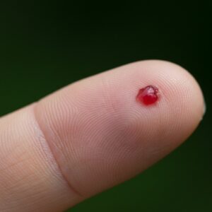
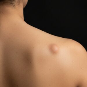
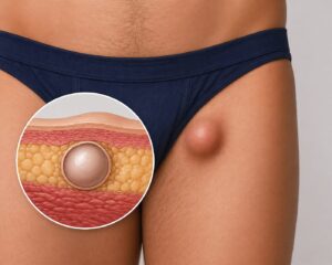
Earlobe keloids can be difficult to manage because they behave differently from standard scars. They tend to thicken over time, respond unpredictably to treatment and often return even after being removed. Many patients explore more than one option before deciding on the right course of action, especially when the keloids have been present for months or years.
At City Dermatology Clinic, patients are given a clear explanation of the benefits and limitations of each approach. The following case-style overview shows how treatment plans are formed and why a stepwise approach is often recommended.
Why Keloids Are Hard to Treat
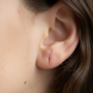
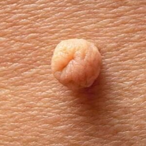
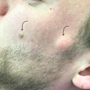
Keloids form when collagen production continues long after a wound has healed. Unlike normal scars, they grow beyond the original area and may become raised, firm or itchy. Earlobes are particularly prone because of piercings, tension on the tissue and the way the ear folds during sleep.
The challenge is that keloids do not behave consistently. Some respond well to medication; others soften briefly but thicken again later. This is why treatment planning involves realistic expectations and a clear understanding of recurrence risk.
First-Line Option: Steroid Injections
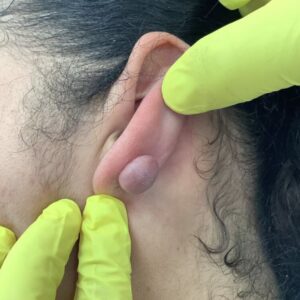
Steroid therapy is often the starting point for small to medium earlobe keloids. It can reduce thickness, soften the scar and make future procedures safer and easier. The steroid used in clinic is typically Kenalog, often mixed with saline and a small amount of local anaesthetic for comfort.
Four earlobe keloids were treated with this method. The injections targeted the densest parts of each scar, aiming to reduce bulk and inflammation. After softening, some keloids improve enough that patients choose to continue with injections alone. Others find the improvement partial and decide to consider surgery once the scar is more manageable.

Patients are counselled on possible side effects, which include skin thinning, colour changes, visible surface vessels and the possibility of needing repeated injections. While steroids can be very effective, they do not cure keloids, they help control them.
Compression clips are used alongside injections, worn continuously to reduce tension on the scar and limit collagen build-up. This combined method can significantly improve results when used correctly.
When Surgery Becomes an Option
If keloids remain bulky after steroid treatment or continue to impact daily life, surgical removal may be considered. This is not a decision taken lightly because ear keloids have one of the highest recurrence rates. With excision alone, recurrence can be anywhere between 60–90%. Even in the best-case scenario, outcomes vary:
• around a third may improve
• around a third may return in a similar form
• around a third may return larger
Because of this, surgery is often planned after steroid injections have softened the scar.
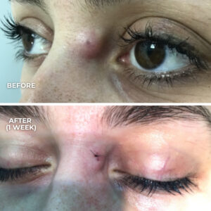
In this case, once the keloids had reduced slightly, the patient opted for intralesional excision, a technique that removes the centre of the keloid while preserving a thin rim of tissue. This reduces tension on the closure and gives the best chance of a stable result.
How Earlobe Keloid Excision Is Performed
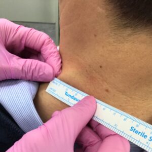
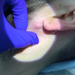
The procedure is done under local anaesthetic, using Lignospan to numb the area. Three keloids were excised intralesionally, with careful removal of scar tissue from within the lesion. Haemostasis is achieved before closing the wound.

Closure is performed with 5/0 nylon sutures, chosen for their precision and minimal skin marking on the delicate earlobe. Micropore tape is used to protect the area after surgery. Once healed, silicone therapy and compression clips help maintain a flatter contour and reduce the chance of recurrence.
Aftercare and Recovery
Post-treatment instructions are designed to protect the wound and manage swelling:
• keep the area dry for 48 hours
• keep tape in place for 4–5 days
• the wound can be showered after 48 hours
• avoid heavy exercise, contact sports and activities that pull on the ear for two weeks
• sutures are removed at around one week
• compression clips should be worn daily as advised
Patients are encouraged to monitor for redness, swelling, increased pain or malodour and contact the clinic if concerned.
Follow-up is an essential part of keloid management. Even after successful excision, the scar continues to remodel for many months. Regular review helps identify early recurrence so it can be treated promptly with steroid top-ups or silicone therapy.
Why a Combined Approach Works Best
Earlobe keloids rarely respond to a single treatment. The best outcomes often come from a tailored approach combining staged steroid injections, careful surgical techniques and strict compression therapy afterwards.
The goal is not only to remove the visible scar but to reduce the biological triggers that caused it to form in the first place. By softening the tissue first, excising conservatively and supporting the wound during healing, the chance of recurrence is significantly reduced compared with surgery alone.
This method helps patients achieve a flatter, more comfortable and more stable result over time, with close monitoring and ongoing guidance throughout the healing process.