Basal cell carcinoma (BCC) is the most common type of skin cancer, but also one of the most treatable when detected early. It grows slowly, rarely spreads to other parts of the body, and can usually be removed completely with the right treatment.
At City Dermatology Clinic in London, our dermatologists regularly diagnose and treat BCCs, using precise techniques to clear the cancer while minimising scarring.
What Basal Cell Carcinoma Is
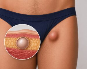
BCC develops in the basal cells, which are found in the lowest layer of the epidermis (the outer layer of the skin). These cells are responsible for creating new skin cells as older ones shed.
When the DNA in these basal cells is damaged, most often by ultraviolet (UV) light from the sun or tanning beds, it triggers abnormal growth, forming a slow-growing tumour.
Why early detection matters
Although BCC is rarely life-threatening, delaying diagnosis can allow the cancer to grow deeper, damaging surrounding skin, nerves, or bone. Early detection means:
- Simpler, less invasive treatment
- Smaller scars
- A lower risk of recurrence
Common Causes and Risk Factors
Several factors can increase the likelihood of developing basal cell carcinoma.
Sun and UV exposure
Cumulative sun exposure is the biggest cause of BCC. This includes years of daily incidental sun exposure, like walking to work, and episodes of intense sunburn.
Skin type, genetics, and age
- Fair skin, light eyes, or freckles
- Family history of skin cancer
- Age over 50, though younger patients are increasingly affected
Other contributing factors
- Long-term use of tanning beds
- Weakened immune system
- Chronic wounds or scarring in the affected area
Early Signs and Symptoms
BCC can look very different from person to person, which is why regular skin checks are important.
Appearance of BCC in different forms
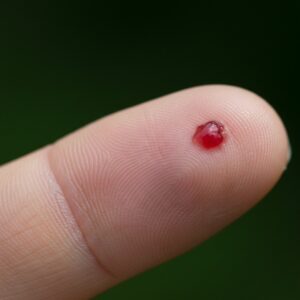
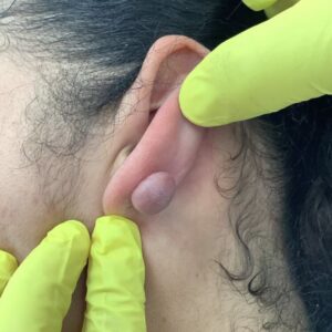
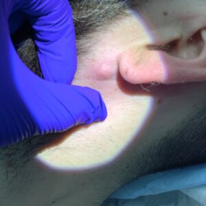
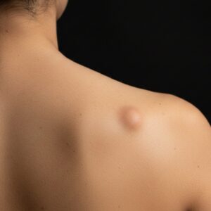
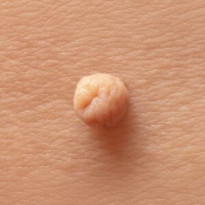
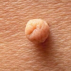
- Nodular BCC: A shiny, flesh-coloured or pink bump that may bleed easily or form a scab.
- Superficial BCC: A flat, red patch that looks like eczema or a rash.
- Morpheaform BCC: A scar-like, waxy patch that may be subtle but invasive under the skin.
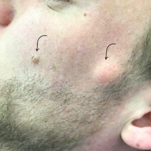
High-risk areas of the body
BCC most often develops in areas that get the most sun exposure, including:
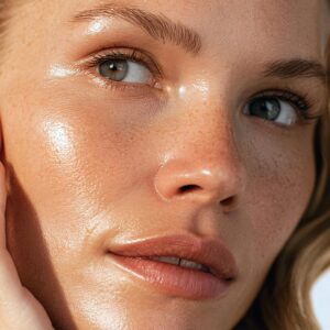
- Face, especially the nose and cheeks
- Ears and scalp
- Neck and shoulders
- Backs of the hands
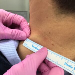
Diagnosis and Assessment
Accurate diagnosis is key to ensuring the right treatment.
Visual checks and dermoscopy
A dermatologist examines the lesion visually and often uses dermoscopy, a magnifying device with polarised light, to analyse deeper skin structures and identify features unique to basal cell carcinoma.
When a biopsy is needed
If there’s any uncertainty or to plan treatment, a skin biopsy is performed. This involves removing a small tissue sample for analysis under a microscope. Biopsies are quick, done under local anaesthetic, and provide a clear diagnosis.
Treatment Options
Basal cell carcinoma treatment depends on the size, depth, and location of the tumour, but the goal is always complete removal with minimal scarring.
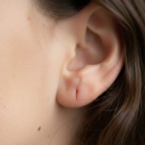
Surgical excision
For most BCCs, the lesion is surgically removed with a small margin of healthy skin to ensure clear boundaries. Stitches are placed for neat healing, and results are reviewed to confirm the tumour is fully cleared.
Mohs micrographic surgery
For facial or high-risk BCCs, Mohs surgery offers the highest cure rate while preserving as much healthy tissue as possible. The tumour is removed in thin layers, with each layer checked under a microscope until no cancer cells remain.
Non-surgical treatments
For smaller or superficial BCCs, other options may be recommended, such as:
- Cryotherapy: Freezing the lesion with liquid nitrogen.
- Topical treatments: Prescription creams containing imiquimod or fluorouracil.
- Photodynamic therapy (PDT): Using light-sensitive medication and a special light source to destroy cancer cells.
Recovery and Monitoring
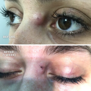
Healing and scar care
- Surgical wounds typically heal within 7–14 days.
- Using silicone gel or tape and protecting the area from sun exposure can improve scar appearance.
- Most patients resume normal activities the next day.
Importance of regular skin checks
Once you’ve had a BCC, you’re at higher risk of developing another. Regular follow-ups with a dermatologist help detect any new lesions early, when treatment is simplest.
Prevention Tips
Daily sun protection
- Use a broad-spectrum SPF 30+ daily, even in cloudy weather.
- Wear hats and UV-protective clothing during prolonged outdoor exposure.
Skin self-checks and when to see a dermatologist
- Look for non-healing spots, shiny bumps, or patches that bleed or scab repeatedly.
- Book an appointment promptly if you notice any changes.