An intradermal nevus is one of the most common types of benign moles. These raised, flesh-coloured or slightly pigmented bumps are usually harmless, but many people choose to have them checked or removed for cosmetic reasons, irritation, or peace of mind.
At City Dermatology Clinic in London, our dermatologists regularly assess and remove intradermal nevi using techniques that are safe, quick, and designed to minimise scarring.
What an Intradermal Nevus Is
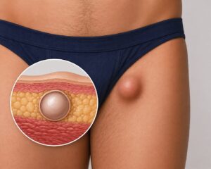
An intradermal nevus is a mole made up of clusters of melanocytes, the cells that produce pigment, located deeper in the dermis layer of the skin.
Definition and how it forms
Most intradermal nevi develop during childhood or adolescence and slowly become more raised with age as pigment cells settle deeper into the skin. They often fade in colour over time, sometimes appearing skin-toned rather than dark brown.
Difference between intradermal and other types of moles
- Junctional nevi: Flat, dark moles where pigment cells are near the surface of the skin.
- Compound nevi: A mix of junctional and intradermal cells, often slightly raised and pigmented.
- Intradermal nevi: Raised, dome-shaped, often lighter in colour, and made up of pigment cells deeper in the skin.
Common Locations and Appearance
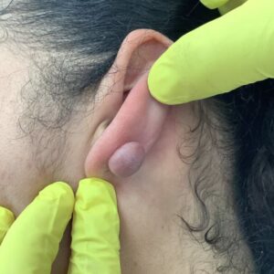
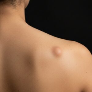
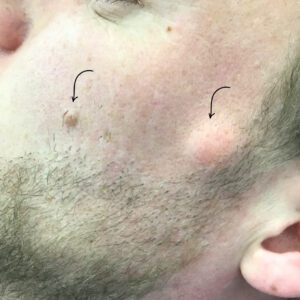
Intradermal nevi can form almost anywhere, but they are most commonly found on:
- The face (particularly cheeks, forehead, and nose)
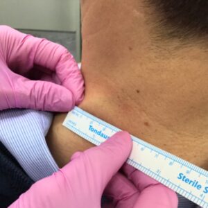
- Neck
- Scalp
- Upper body
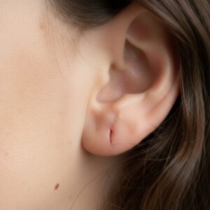
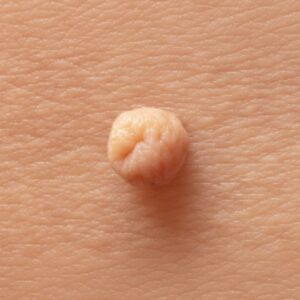
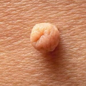
Features that make intradermal moles easy to identify
- Raised, dome-shaped surface
- Skin-coloured, pink, or light brown
- Smooth texture, sometimes with a hair growing from it
- Stable appearance that doesn’t change rapidly
Are Intradermal Nevi Dangerous?
The good news is that intradermal nevi are almost always benign. They rarely, if ever, transform into melanoma or other forms of skin cancer.
Risk of becoming cancerous
The risk of cancerous change in an intradermal nevus is extremely low. However, any mole that changes in size, shape, or colour, or begins bleeding or itching, should always be assessed by a dermatologist to rule out malignancy.
When changes should be checked by a dermatologist
Seek medical advice if you notice:
- Rapid growth or change in colour
- Irregular or jagged edges
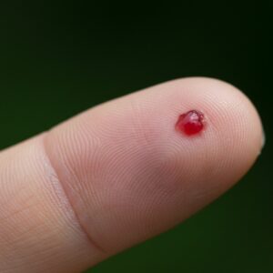
- Persistent bleeding or ulceration
- New pain, itching, or irritation
Diagnosis
Most intradermal nevi can be confidently diagnosed during a routine dermatology consultation.
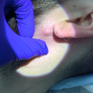
Visual examination and dermoscopy
A dermatologist will examine the mole visually and often use a dermatoscope, a small magnifying device with polarised light, to look at the deeper structures of the mole. This helps differentiate a harmless intradermal nevus from atypical moles or early melanoma.
When a biopsy may be recommended
If the mole looks unusual, has changed recently, or shows irregular features under dermoscopy, a biopsy may be performed. This involves removing a small sample or the entire mole for microscopic analysis.
Treatment and Removal Options
Intradermal nevi don’t need to be removed unless they are:
- Causing irritation or catching on clothing
- Located in a cosmetically sensitive area
- Changing in appearance or causing concern
At City Dermatology Clinic in London, our dermatologists offer several safe and effective removal options.
Shave excision
This is the most common method for raised, benign moles. The mole is numbed with local anaesthetic, then carefully shaved level with the surrounding skin. This is quick, minimally invasive, and leaves only a fine mark that fades with time.
Surgical excision
For deeper or more complex intradermal nevi, a surgical excision may be recommended. The mole and its root are removed entirely, and the skin is closed with fine stitches for optimal healing. This method ensures complete removal and is often used if there’s any diagnostic uncertainty.
Cosmetic vs. medical reasons for removal
- Cosmetic: For smooth, flatter skin or to remove visible moles from areas like the face.
- Medical: When there are changes in the mole or if it’s frequently irritated or injured.
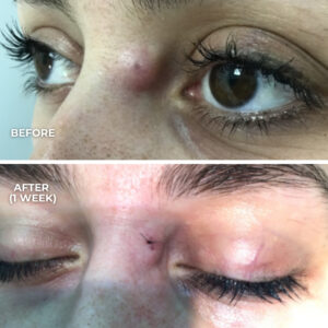
Healing, Recovery & Scarring
Healing after removal is straightforward and usually involves minimal downtime.
What to expect after removal
- Shave excision sites form a small flat scab that heals within 7–10 days.
- Surgical excision sites may have stitches removed after 7–14 days, depending on the area.
Minimising scars for the best cosmetic results
- Keep the area clean and protected while it heals.
- Apply silicone gel or tape once the wound has closed to flatten and fade scars.
- Use sun protection daily to prevent pigmentation or darkening of the healing site.
When to See a Dermatologist
Regular mole checks are the safest way to ensure your skin remains healthy. Book a consultation if you notice:
- Rapid changes in a mole’s appearance
- New moles developing after the age of 30
- Moles that bleed, crust, or become painful
At City Dermatology Clinic, our dermatologists — including Dr Parviz Sadigh and Dr Jana Torres-Grau, provide expert assessments and safe, effective removal options tailored to your needs.