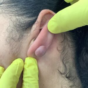
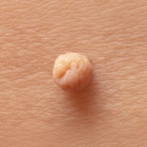
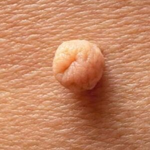
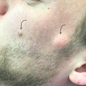
A patient presented with a seven-month history of multiple keloid scars affecting both ear helices. There were two small keloids on the right helix and one on the left, each measuring under 1 cm. They were raised, firm and causing discomfort, especially when wearing in-ear headphones or sleeping on the side. No sinister features were noted on examination, and the patient was keen on surgical removal.
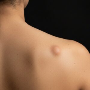
Ear keloids form when the skin heals with excess scar tissue, often after piercing, minor trauma or inflammation. Although small, they can be persistent and tend to grow slowly. Excision with targeted closure is one of the most effective approaches for well-defined, early keloids, particularly when combined with precise wound care to reduce the chance of recurrence.
The following explains how helix keloid removal is performed, what patients can expect during treatment and how we manage scarring to support long-term results.
Assessment of Helical Keloid Scars
Keloids on the ear helix behave differently from keloids elsewhere on the body. The cartilage and contour of the ear create tension lines that can influence how the scar forms and grows. Even small keloids can cause discomfort because of the ear’s shape and the way it interacts with sleep position, headphones or daily movement.
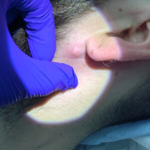
During the consultation, Dr Parviz Sadigh assessed three distinct lesions. All were under 1 cm, raised and well-defined. Their size and shape made them suitable for intralesional excision, which involves removing the keloid from within its borders while preserving normal surrounding tissue. This technique reduces the bulk of the scar while avoiding unnecessary tension on the ear.
Because no concerning features were seen on examination, surgical treatment was agreed as the first-line option, with a discussion about recurrence risk and the importance of aftercare.
How Intralesional Excision Works
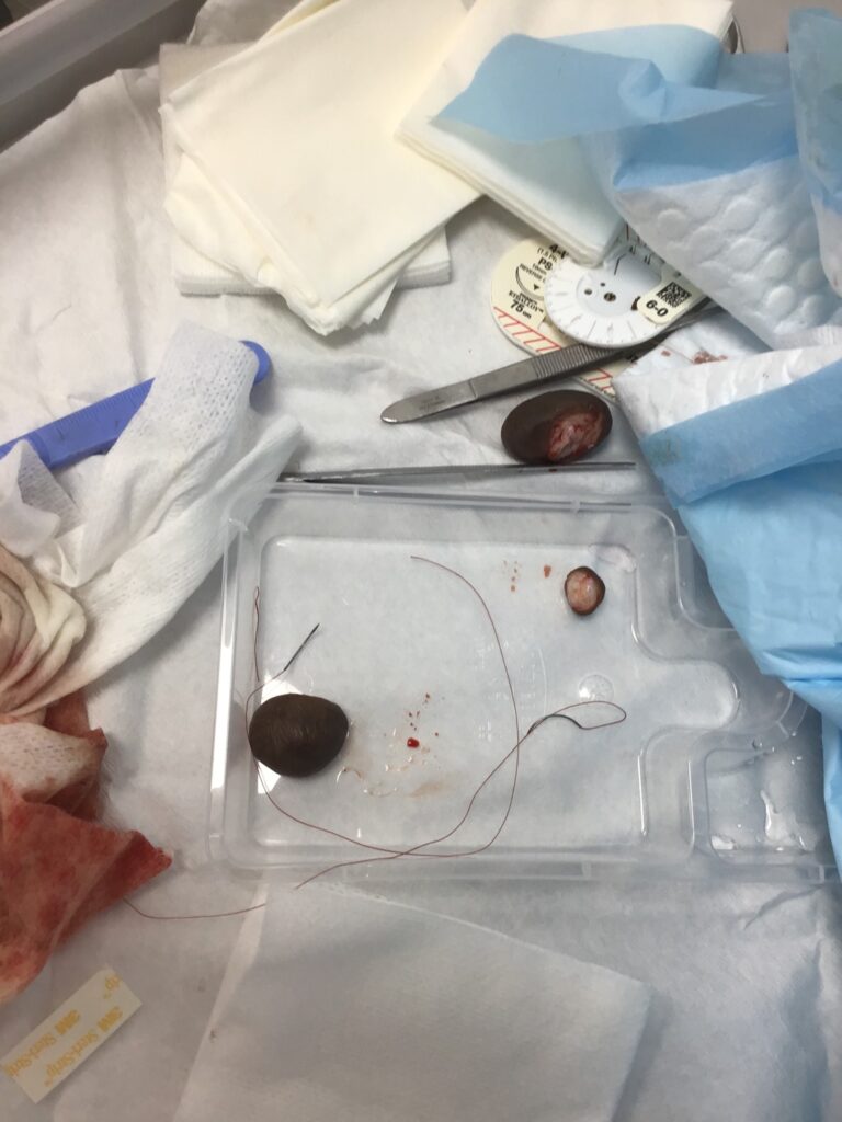
Intralesional excision focuses on removing the core of the keloid without creating a wide external wound. Instead of cutting around the entire edge of the scar, the surgeon excises the thicker central portion while preserving a thin rim of tissue. This approach is particularly suitable for small helix keloids because it reduces tension during closure, which is a major factor in preventing keloids from reforming.
The patient was treated under local anaesthetic using Lignospan LA, ensuring full numbness throughout the procedure. Each keloid was carefully excised from within its borders. Once the bulk of the lesion was removed, the base was smoothed and the wound bed prepared for closure.
This technique is widely used for early-stage ear keloids, especially those related to piercings. When performed precisely, it offers a strong balance between cosmetic outcome and long-term stability.
Surgical Technique and Closure
After each keloid was removed, haemostasis was achieved to ensure a clean field. Dr Sadigh used a layered closure approach tailored for the delicate structure of the ear. The deeper layer was sutured with 4/0 Vicryl Rapide, which supports the wound internally as it heals. The skin layer was refined with 6/0 Rapide, a finer suture chosen to minimise visible marking on the helix.

The aim of closure in ear keloid surgery is to create a flat, even contour without tension. Because the ear moves and bends, any stress on the wound can increase the chance of excessive scar tissue returning. By using dissolvable sutures in controlled layers, the wound heals with support but without the bulk of long-lasting stitches.
An Opsite spray dressing was applied over the sites to protect them during the initial healing phase. This forms a thin, breathable layer that shields the incision while allowing the patient to go about their day normally.
What Patients Can Expect During the Procedure
The treatment is carried out under local anaesthetic, so the patient remains comfortable throughout. Most procedures take around 20–30 minutes depending on the number and size of keloids. The patient typically feels only light pressure once the area is numb.
Ear keloid excision is a delicate procedure, but downtime is minimal. Most patients return to work or normal routine shortly afterward. Because the lesions in this case were under 1 cm, the incisions were small and closed neatly, requiring only basic aftercare.
Healing and Aftercare
Healing after ear keloid removal usually progresses steadily as long as the wound is protected. The ear should be kept dry for the first day, and care should be taken when sleeping or using headphones to avoid unnecessary pressure. The Opsite spray remains in place as a protective barrier, and once it naturally lifts or is removed as instructed, patients may transition to light wound care.
Keloids can recur, so long-term management is an important part of the plan. Patients may be advised to use silicone gel or silicone sheeting after the wound has fully sealed. These methods help flatten the scar and reduce collagen overproduction. Follow-up is arranged to assess healing and discuss additional treatments such as steroid injections if required.
Our Expertise in Keloid Treatment
City Dermatology Clinic treats keloids of all sizes, from small helix lesions to larger scars caused by trauma or surgery. Dr Parviz Sadigh performs these procedures regularly and uses techniques that prioritise precision, comfort and long-term stability. Treatment plans are personalised to reduce recurrence risk and support smooth cosmetic results, with clear guidance at every stage.