Healing after mole removal happens in stages and varies depending on the removal method, location of the mole, and individual skin healing. This guide explains what patients typically see during recovery and how healing differs between common mole removal techniques.
Images are often helpful for reassurance, but it is important to understand that healing timelines and appearance vary between individuals.
Why Healing Looks Different for Each Patient
Healing after mole removal is not identical for everyone. Even when the same technique is used, the appearance and speed of healing can vary.
Factors that influence healing include:
- Removal technique
Shave excision, surgical excision, electrocautery, laser treatment, and cryotherapy affect different skin layers and heal in different ways. - Mole size and depth
Deeper or larger moles usually take longer to heal and may leave a more noticeable mark. - Body location
Areas under tension or movement (such as the back, shoulders, or joints) often heal more slowly than areas with thinner skin, such as the face. - Individual skin type and healing response
Skin thickness, pigmentation, and tendency to scar differ between individuals. - Aftercare and wound protection
Following aftercare advice and avoiding trauma or friction helps support normal healing.
Because of these variables, healing images should be used as a general guide only, not a prediction of outcome.
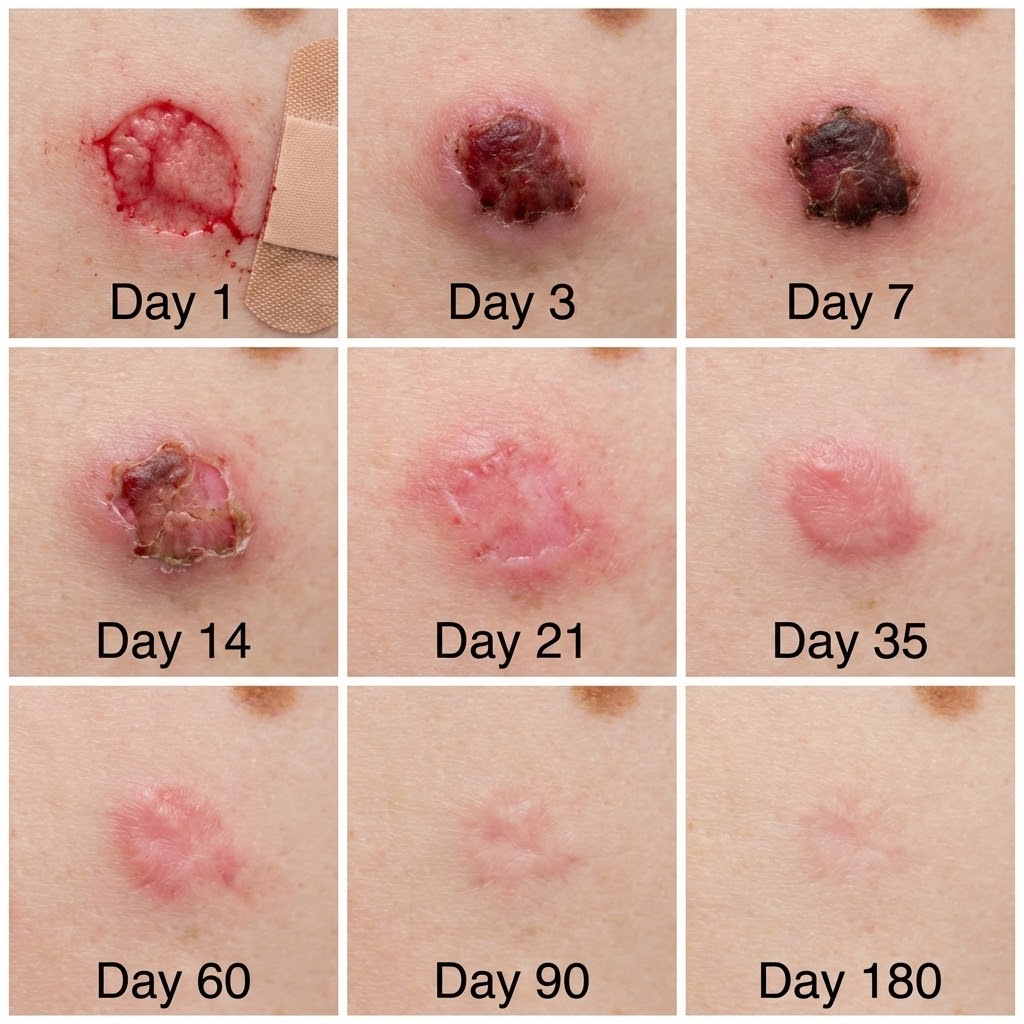
General Mole Removal Healing Timeline
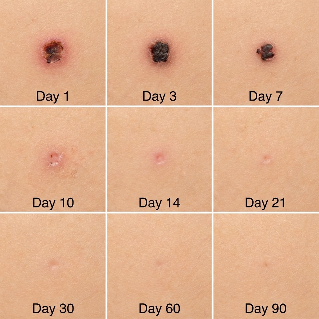
The table below outlines the typical stages of healing following mole removal.
Timeframes are approximate and vary depending on the procedure and individual healing response.
| Healing Stage | Typical Timeframe | What You May Notice | What This Means |
|---|---|---|---|
| Immediate phase | Day 0–3 | Redness, mild swelling, small amount of oozing, dressing or scab present | Normal early response as the skin seals and inflammation begins |
| Early repair | Day 4–10 | Scab darkens and dries, redness gradually reduces | New skin is forming underneath the scab |
| Surface healing | Week 2–4 | Scab falls away naturally, pink or pale skin visible | Skin surface has closed; area may still appear sensitive |
| Scar maturation | Week 4–12+ | Pinkness fades, scar softens and flattens | Healing continues beneath the surface and appearance gradually settles |
Important notes:
- Stitches, if used, are usually removed within 7–14 days, depending on location.
- Pink or red skin during early healing is common and does not indicate infection.
- Final scar appearance can continue to change for several months.
If healing appears delayed or symptoms worsen rather than improve, a clinician can assess the area and advise appropriately.
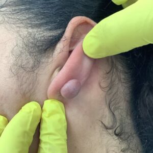
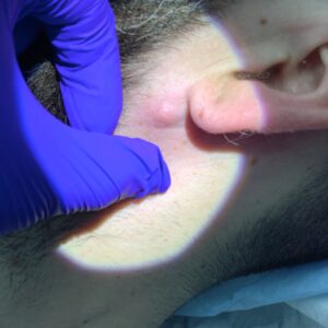
Shave Excision Healing Stages
Shave excision involves removing a raised or dome-shaped mole at skin level under local anaesthetic. Because stitches are not usually required, healing occurs from the surface downward rather than through a closed incision.
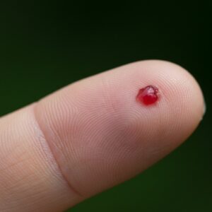
In the first few days, the treated area typically appears red and slightly raw, with a shallow wound where the mole was removed. A scab usually forms within 24 hours. Mild oozing or spotting can occur initially and is part of normal early healing.
Over the following one to two weeks, the scab dries and gradually darkens as the skin underneath repairs itself. During this stage, the area may feel tight or itchy. It is important not to pick the scab, as this can delay healing and increase the risk of scarring.
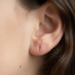
Once the scab separates naturally, new skin becomes visible. This skin is often pink or lighter than the surrounding area and may appear slightly indented. This is a normal part of the healing process and does not indicate a problem.
Over the next several weeks, the colour of the new skin usually fades and blends more with the surrounding skin. In some cases, a flat, faint mark remains. The final appearance depends on individual healing response, skin type, and the size and depth of the original mole.
Histology can be arranged following shave excision when clinically appropriate, and healing progress can be reviewed during follow-up if there are any concerns.
Surgical Excision Healing Stages
Surgical excision is used when a mole requires full removal, including deeper tissue, and is performed under local anaesthetic. The wound is closed with stitches, so healing involves both surface skin repair and deeper tissue recovery.

In the first few days after the procedure, the area is usually red, slightly swollen, and may feel tight or tender. A dressing is typically applied, and mild bruising can occur, particularly on areas such as the trunk or limbs. These early changes are part of the normal inflammatory phase of healing.
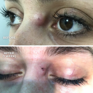
During the first one to two weeks, the wound edges begin to seal and the surrounding redness gradually reduces. Stitches are usually removed within this period, depending on the body location and wound tension. It is common for the incision line to look raised or pink at this stage.
After stitch removal, the surface skin continues to strengthen. The scar may remain pink, firm, or slightly lumpy for several weeks. This reflects normal collagen remodelling beneath the skin and does not indicate poor healing.
Over the following months, the scar typically softens and becomes flatter and less noticeable. Colour changes gradually fade, although the final appearance varies between individuals. Factors such as skin type, location, and individual scarring tendency all influence long-term results.
Surgical excision allows for routine histological analysis of the removed tissue. If there are concerns about healing, scar appearance, or symptoms such as increasing pain or redness, a clinician can review the area and provide guidance.
Electrocautery Healing Stages
Electrocautery is sometimes used to remove very small, superficial moles or pigmented lesions. The technique uses controlled heat to destroy the lesion at the skin surface and seal small blood vessels at the same time. Stitches are not required.
Immediately after treatment, the area usually appears red with a small, dry wound or shallow crater. Because the tissue is cauterised, bleeding is minimal. Mild soreness or warmth around the area can occur on the first day.
Within a few days, a firm scab forms over the treated site. This scab protects the underlying skin while healing takes place. It is common for the area to look darker or slightly crusted during this phase.
Over the following one to two weeks, the scab gradually dries and separates naturally. New skin forms underneath and may appear pink or lighter than the surrounding skin. Temporary redness is expected and usually settles with time.
In the weeks that follow, the skin continues to mature and blend with the surrounding area. In some cases, a small flat mark remains. Healing speed and final appearance vary depending on the size of the lesion, skin type, and individual healing response. Histological analysis is not always possible with electrocautery, which is considered during assessment.
Laser Treatment Healing Appearance
Laser treatment targets pigment in the skin and is sometimes used for selected superficial pigmented lesions. It affects the surface layers of the skin rather than removing the mole in one piece.
Immediately after laser treatment, the area may appear red or slightly swollen. A mild burning or warm sensation can occur for a short time. Some lesions darken or crust over within the first 24 to 48 hours.
During the first one to two weeks, light crusting or scabbing may develop and then shed naturally. As the surface heals, the treated area may look pink or temporarily lighter or darker than the surrounding skin.
Over the following weeks, skin colour usually continues to even out. Any residual redness gradually fades as the skin recovers. Final appearance varies and can take several weeks to settle fully.
Laser treatment does not remove deeper mole tissue and does not allow for histological examination. For this reason, it is not suitable for all moles and is only considered following clinical assessment.
Cryotherapy Healing Stages
Cryotherapy uses controlled freezing to destroy superficial skin lesions by causing targeted damage to the treated tissue. It is sometimes used for selected benign lesions but is not suitable for all moles.

Immediately after treatment, the area may appear red and swollen. A stinging or burning sensation can occur for a short period following freezing. In some cases, a blister forms within the first 24 to 48 hours, particularly if deeper freezing was required.
Over the next few days, any blister usually dries out and a scab forms. The treated area may look darker or crusted during this phase. This is part of the normal healing process as damaged tissue separates from the skin.
Within one to two weeks, the scab gradually falls away on its own. New skin becomes visible underneath and is often pink or lighter than the surrounding area. Temporary changes in skin colour are common at this stage.
Over the following weeks, the skin continues to heal and settle. Colour and texture usually improve gradually, although healing time and final appearance vary between individuals. Cryotherapy does not remove deeper pigment cells and does not allow for histological analysis, which is considered during clinical assessment.
What Is Not Normal During Healing
Most mole removal sites heal without complication, but certain changes are not expected and should be reviewed.
Signs that warrant clinical assessment include:
- Increasing pain after the first few days rather than gradual improvement
- Redness that spreads beyond the immediate treatment area
- Warmth, swelling, or throbbing around the wound
- Discharge, pus, or an unpleasant odour
- The wound reopening or bleeding that does not settle
- Fever or feeling generally unwell
These features are uncommon but should not be ignored. A clinician can assess whether healing is delayed or if treatment is required.
Will Mole Removal Leave a Scar?
Any procedure that breaks the skin can result in a scar. The appearance of a scar varies between individuals and depends on several factors.
These include:
- The removal method used
- The size and depth of the mole
- Body location (for example, chest and back scars often mature differently to facial scars)
- Individual skin type and scarring tendency
- Wound care and protection during healing
Scars often appear pink or firm in the early weeks and gradually soften and fade over time. A clinician will usually discuss scar expectations before treatment.
When Healing Takes Longer Than Expected
Healing may take longer in certain situations, including:
- Larger or deeper moles
- Areas under tension or frequent movement (such as shoulders, back, or joints)
- Repeated friction from clothing or activity
- Delayed scab separation
- Individual factors such as slower wound healing or scarring tendency
In some cases, follow-up appointments are arranged to monitor healing or provide reassurance.
Final Notes on Healing Stage Pictures
Healing stage pictures are intended to show common patterns, not guaranteed outcomes. They cannot account for individual skin response, body location, or treatment method.
It is normal for healing to look different from images found online or from other patients. If there is uncertainty about whether healing is progressing as expected, a clinician can assess the area and provide guidance.