Cysts under the skin are common, usually benign lumps that can vary in size, location, and appearance. People often search for pictures to understand what a cyst might look like and how it can differ from other skin lumps.
Our images on this page show typical examples of cysts beneath the skin. They are provided for general reference only. A visual comparison cannot confirm a diagnosis, as many skin conditions can look similar. A clinical assessment is required to determine the nature of any lump.
What Do Cysts Under the Skin Usually Look Like?
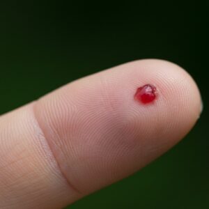
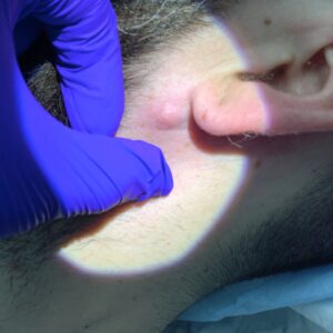
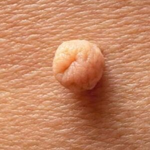
Cysts under the skin often appear as:
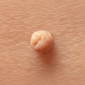
- A round or oval lump beneath the surface
- A swelling that feels smooth or firm to touch
- Skin over the lump that usually looks normal
- A lump that may move slightly under the skin
Some cysts remain small for years, while others gradually increase in size. Growth is usually slow.
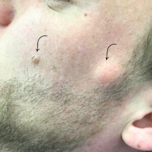
Common Types of Cysts Shown in Pictures
Images of cysts under the skin commonly include:
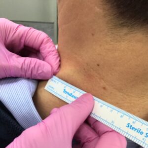
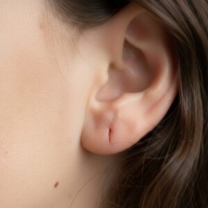
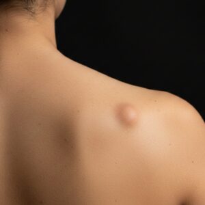
- Epidermoid cysts – often found on the face, neck, or trunk
- Pilar cysts – more common on the scalp
- Sebaceous-type cysts – a term people often use to describe epidermoid cysts
- Inflamed cysts – which may appear red, swollen, or tender
Different cyst types can look similar in photographs, which is why images alone are not enough to identify them.


Why Pictures Are Not Enough for Diagnosis
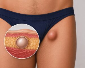
Other conditions – such as lipomas, enlarged lymph nodes, or abscesses – can resemble cysts in pictures. The feel of the lump, its behaviour over time, and its location are just as important as how it looks.
A clinician considers:
- How long the lump has been present
- Whether it is growing or changing
- Whether it is painful or inflamed
- Its position and depth under the skin
When to Consider Medical Assessment
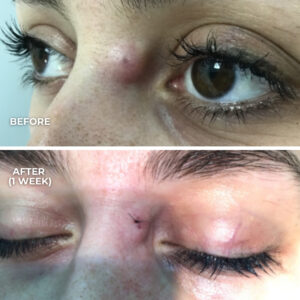
Pictures can be a useful starting point, but cyst removal assessment is usually recommended if a lump:
- Is increasing in size
- Becomes painful, red, or inflamed
- Starts to discharge
- Causes uncertainty or concern
A consultation allows a clinician to examine the area properly and explain whether the lump is likely to be a cyst and what options, if any, may be appropriate.