A cyst on the ear is usually a harmless, slow-growing lump that forms beneath the skin. It can appear on the earlobe, behind the ear, or on the outer ear cartilage. While many ear cysts are painless and stable, some become inflamed, uncomfortable, or cosmetically concerning.
Cyst removal is not always necessary. The first step is assessment to confirm the diagnosis and determine whether treatment is appropriate. This guide explains the common types of ear cysts, why they develop, and what removal involves in a private UK clinical setting.
What is a cyst on the ear?
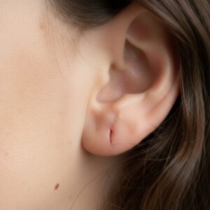
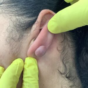
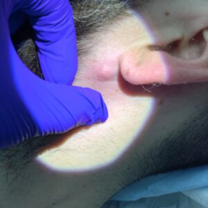
A cyst on the ear is a common, non-cancerous lump that develops beneath the surface of the skin. It may appear on the earlobe, behind the ear, along the outer rim of the ear (pinna), or occasionally near the ear canal entrance.
Most ear cysts grow slowly and are not painful. They often feel smooth, rounded and mobile under the skin. Some remain unchanged for years. Others may gradually increase in size or become inflamed.
At City Dermatology Clinic, ear lumps are assessed as part of a structured dermatology or minor surgery consultation. Although many ear lumps are cysts, other conditions can present in a similar way, including:
- Lipomas (fatty lumps)
- Keloid scars, particularly after piercings
- Enlarged lymph nodes behind the ear
- Benign skin growths
Because the ear contains cartilage and has relatively thin skin in certain areas, accurate identification is important before considering removal. A clinical examination allows the clinician to confirm whether the lump is likely to be a cyst and whether treatment is appropriate.
Assessment is particularly important if the lump:
- Has changed in size
- Has become painful or red
- Has started to discharge
- Has an unusual appearance
A consultation provides clarity and helps determine next steps without assuming removal is required.
Types of cysts that can occur on the ear
Several types of cyst may develop on or around the ear. Understanding the likely type helps guide management.
Epidermoid cyst
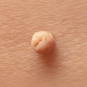
This is the most common type seen on the ear. Epidermoid cysts form when skin cells become trapped beneath the surface rather than shedding normally. Over time, these cells accumulate and create a small sac filled with keratin (a soft, cheese-like material).
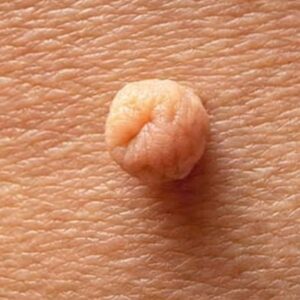
They often present as:
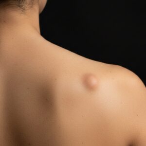
- Firm, dome-shaped lumps
- Skin-coloured or slightly yellow
- Slow-growing
Epidermoid cysts can develop on the earlobe, especially in areas affected by piercings or minor trauma.
Pilar (trichilemmal) cyst
Pilar cysts are more frequently found on the scalp but can occur near the ear. They tend to feel firmer than epidermoid cysts and may be less likely to rupture spontaneously.
“Sebaceous cyst” (common patient term)
Many patients describe an ear lump as a sebaceous cyst. In clinical practice, most of these are epidermoid cysts. True sebaceous cysts, arising from sebaceous glands, are less common.
Inflamed or infected cyst
A cyst can become inflamed or infected. Signs may include:
- Redness
- Swelling
- Tenderness
- Warmth
- Discharge
When inflamed, the lump may increase rapidly in size and become painful. In this situation, immediate surgical removal is not always appropriate. The inflammation may need to settle first before definitive excision can be considered.
At assessment, the clinician evaluates not only the type of cyst but also its size, location and current state (calm versus inflamed), as this affects management decisions.
Why people seek assessment or removal
Many ear cysts are medically harmless. However, patients commonly request assessment for practical or personal reasons.
Cosmetic concerns
The ear is a visible area. Lumps on the earlobe or outer ear can be noticeable, particularly if they enlarge. Even small cysts may cause self-consciousness.
Discomfort
Cysts located behind the ear or on the upper ear can rub against:
- Glasses
- Headphones
- Helmets
- Face masks
Repeated pressure may lead to irritation or tenderness.
Recurrent inflammation
Some cysts repeatedly become inflamed. This can cause intermittent pain, redness and discharge. Patients who have experienced repeated flare-ups often request removal to reduce the likelihood of further episodes.
Uncertainty about diagnosis
Not all lumps are cysts. Patients frequently attend because they are unsure what the lump represents. In our booking data, assessment for lumps and lesions forms a significant proportion of consultations, reflecting this diagnostic uncertainty. A structured consultation allows the clinician to examine the area and clarify whether it is likely benign.
Previous infection or drainage
If a cyst has previously been drained but not fully excised, recurrence can occur. In such cases, removal of the cyst lining may be discussed to reduce the chance of further recurrence, although recurrence is still possible in some instances.
Assessment and treatment options
Assessment at City Dermatology Clinic is consultation-led. The first step is confirming that the lump is likely to be a cyst and not another type of skin lesion.
During the consultation, the clinician will:
- Examine the ear and surrounding skin
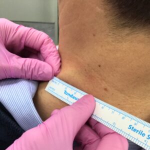
- Assess the size, depth and mobility of the lump
- Check for signs of inflammation or infection
- Discuss symptoms and duration
- Explain whether removal is appropriate
If the lump has atypical features, further evaluation may be recommended. In most cases, however, ear cysts can be assessed clinically.
When removal may be considered
Removal may be discussed if the cyst:
- Is growing
- Is repeatedly inflamed
- Causes discomfort
- Is cosmetically bothersome
- Creates uncertainty about diagnosis
Surgical excision
The most common treatment for an ear cyst is surgical excision under local anaesthetic. This involves:
- Numbing the area
- Making a small incision
- Removing the cyst and its lining (capsule)
- Closing the wound with stitches
Removing the entire lining reduces the likelihood of recurrence. However, recurrence is still possible in some cases.
Inflamed or infected cysts
If the cyst is acutely inflamed or infected, immediate full excision may not be suitable. In these cases:
- Antibiotics may be prescribed if clinically indicated
- Drainage may be considered if there is an abscess
- Definitive removal may be delayed until inflammation settles
This staged approach can reduce complications and improve healing.
Histology
If there is any uncertainty about the diagnosis, the removed tissue may be sent for histological analysis. This is discussed during consultation.
Same-day removal may be possible depending on assessment findings and scheduling, but it is not guaranteed.
Treatment steps and what to expect
Ear cyst removal is usually performed as a minor outpatient procedure.
| Step | What happens |
|---|---|
| Consultation | The cyst is assessed and suitability for removal confirmed. |
| Anaesthetic | Local anaesthetic is injected to numb the area. |
| Excision | A small incision is made and the cyst and lining are removed. |
| Closure | The wound is closed with stitches. |
| Dressing | A protective dressing is applied. |
| Follow-up | A wound check or suture removal may be arranged. |
Procedure time
The procedure itself is typically brief, although exact duration depends on cyst size and location.
Aftercare
Patients are given aftercare advice, which may include:
- Keeping the area clean and dry
- Avoiding unnecessary pressure on the ear
- Monitoring for signs of infection
If stitches are used, they are usually removed at a follow-up appointment unless dissolvable sutures are placed.
Healing time varies between individuals. The ear’s thin skin and cartilage structure mean careful aftercare is important.
Risks, healing and scarring
As with any procedure that breaks the skin, ear cyst removal carries risks. These are generally minor but should be understood in advance.
Possible risks include:
- Bleeding
- Infection
- Wound separation
- Recurrence of the cyst
- Scarring
Scarring on the ear
The ear, particularly the earlobe, can be prone to noticeable scarring in some individuals. Patients with a history of keloid or hypertrophic scarring should inform the clinician during consultation.
Any procedure that involves an incision will leave a scar. The final appearance depends on:
- Individual healing response
- Cyst size
- Exact location
- Surgical technique
- Aftercare adherence
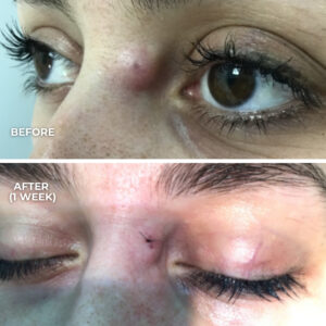
Scars typically mature and soften over several months.
Pain and recovery
Mild discomfort is common in the first few days. Pain levels vary between individuals. Most patients can resume normal daily activities shortly after the procedure, although pressure or friction on the ear should be avoided during early healing.