Searches for mole removal before and after and facial cyst removal often reflect a need for clarity rather than cosmetic comparison. Patients want to understand what assessment involves, how procedures are planned, and what changes to expect over time, particularly when lesions are on visible areas such as the face or hairline.
Recent facial procedures provide a useful clinical context for explaining how “before and after” should be interpreted in medical practice.
Assessment Comes Before Treatment
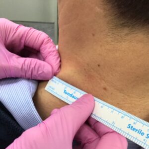
Before any mole or cyst removal, a skin check is performed. This involves assessing the lesion itself as well as the surrounding skin, taking into account size, location, appearance, and patient history. Even when a lesion appears benign, assessment helps determine whether removal is appropriate and which technique may be suitable.
In some cases, clinicians may also discuss alternative approaches such as shave removal or biopsy, particularly if there is diagnostic uncertainty or if histological examination may be required. These decisions are made on an individual basis and form an important part of the “before” stage.
Facial Lesions: Why Technique Matters
The face presents specific considerations. Skin tension lines, proximity to important structures, and cosmetic visibility all influence surgical planning. As a result, technique selection and closure methods are carefully chosen to balance complete removal with controlled wound healing.
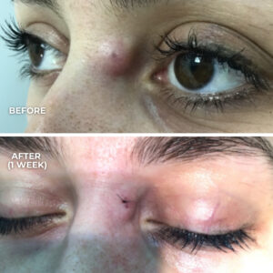
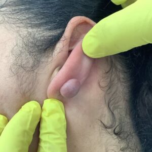
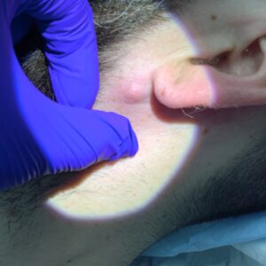
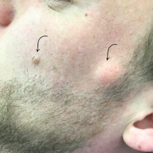
In one recent case, two facial lesions were managed during the same clinical episode:
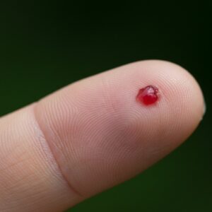
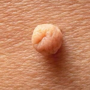
- A benign mole on the left cheek, measuring approximately 1.2 mm
- A cyst on the left temple, extending across the hairline in the zygomatic region
Both lesions were assessed prior to removal to confirm suitability and to plan incision placement and closure.
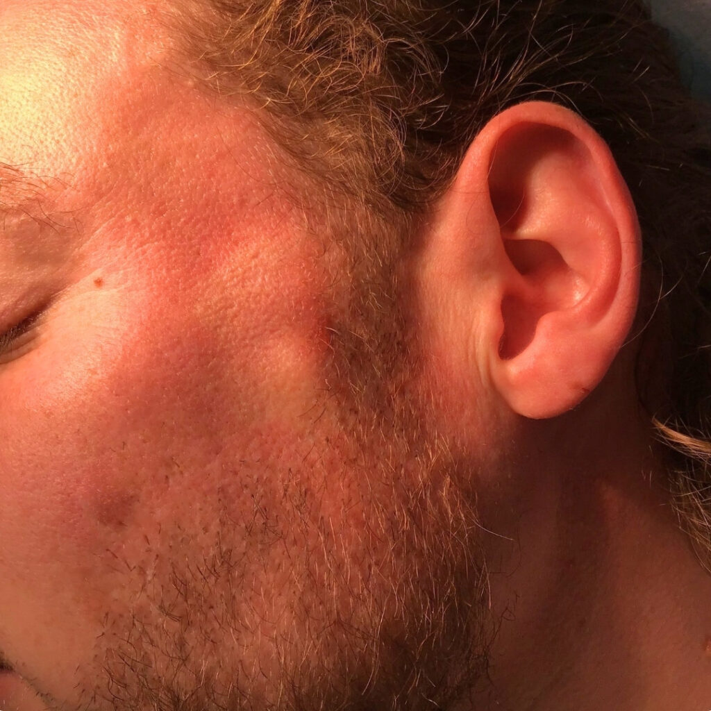
The mole on the left cheek was removed by surgical excision and closed using 6/0 Nylon sutures, a fine suture material commonly used on facial skin to support accurate edge approximation.
The cyst on the left temple was removed through a minimal approach within the hairline, reducing visible disruption to surrounding skin. Closure was again performed using 6/0 sutures, reflecting the need for precision in this area.
These procedures were performed by Mr Georgios Pafitanis, with assessment and technique selection guided by lesion type and anatomical location.
What “After” Means in Clinical Terms
In medical practice, “after” refers to more than immediate appearance. It includes:
- Early wound healing and dressing care
- Suture management and follow-up
- Monitoring for infection or delayed healing
- Review of histology where applicable
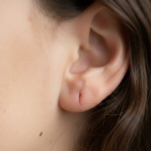
Facial skin often heals well, but scarring is always possible, and its appearance varies between individuals. Factors such as skin type, healing response, sun exposure, and aftercare all influence the final result.
Setting Realistic Expectations
Before-and-after images can be helpful, but they should be interpreted cautiously. They represent individual cases rather than guaranteed outcomes. No two lesions, patients, or healing processes are the same.
For this reason, clinicians focus on assessment-led decision-making, clear explanation of options, and realistic discussion of healing and scarring. The goal is informed understanding rather than comparison.
Transparency Through Education
Publishing educational content around mole and cyst removal helps demystify minor skin surgery and supports patient understanding. These explanations are not intended to encourage treatment, but to clarify what assessment and removal typically involve when lesions are on visible areas of the face.