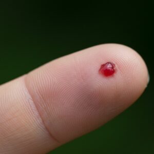
Not all moles require treatment, but some changes can understandably cause concern. A mole that looks different, has altered over time, or has begun to cause symptoms often prompts patients to seek specialist advice, not necessarily because something is wrong, but because reassurance or clarity is needed.
Assessment of suspicious moles is based on careful clinical evaluation rather than appearance alone. This includes understanding how the mole has behaved over time, examining it with specialist tools, and deciding whether monitoring, removal, or no further action is appropriate. In many cases, removal is advised to confirm diagnosis or address ongoing concern, while in others reassurance is all that is required.
As part of City Dermatology Clinic, our doctors manage both clinically indicated mole removals and cosmetic mole removal, with dermatologist-led assessment and access to plastic surgeons when surgical planning or cosmetic considerations are relevant. This specialist, assessment-first approach allows decisions to be tailored to the individual mole and patient, rather than following a routine pathway.
What Is Considered a “Suspicious” Mole?
In clinical terms, a mole is considered suspicious when it shows features that justify closer medical assessment. This does not mean that a diagnosis has been made or that cancer is present. Rather, it indicates that the mole does not fully fit the pattern of a typical, stable benign lesion and warrants professional review.
Moles can change for many reasons. Some changes are harmless and relate to ageing, hormonal shifts, or cumulative sun exposure. Others may reflect underlying cellular changes that need to be assessed more carefully. The purpose of identifying a mole as suspicious is therefore not to alarm, but to determine whether further evaluation or removal is appropriate.
From a specialist perspective, the term “suspicious” is deliberately cautious. It acknowledges uncertainty and triggers a structured assessment rather than an assumption about outcome. This distinction is important, as many patients understandably associate the word with worst-case scenarios, when in reality most assessed moles are benign.
Changes Specialists Look For During Assessment
When assessing a mole, clinicians consider more than a single visual feature. Assessment is based on a combination of appearance, behaviour over time, symptoms, and clinical context.
Key features that may prompt closer evaluation include:
- Change over time
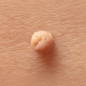
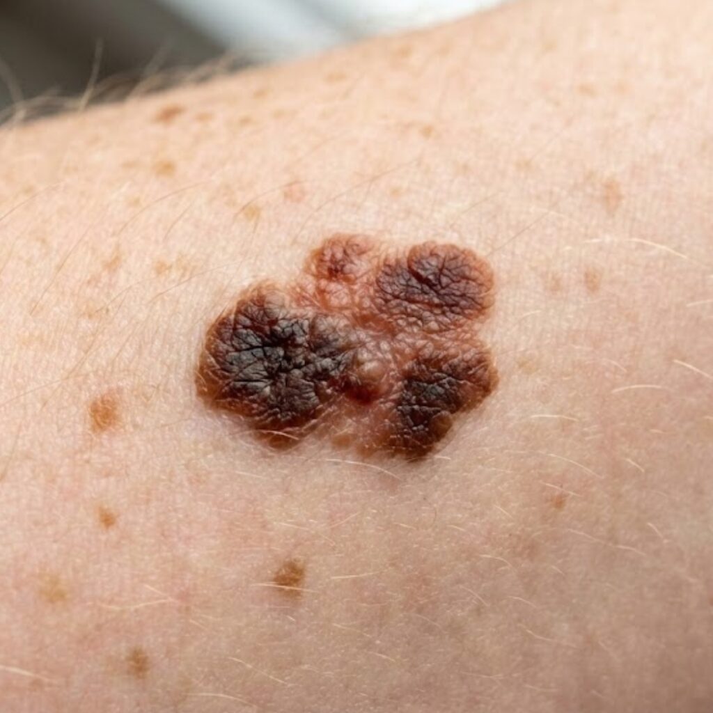
Alterations in size, shape, colour, or thickness are often more relevant than how a mole looks at a single point in time. A mole that has evolved recently may merit assessment even if it appears relatively small. - Irregular structure or colour pattern
Moles with uneven borders, multiple shades of colour, or asymmetry may be examined more closely, particularly if these features are new. - New symptoms
Sensations such as itching, tenderness, bleeding, crusting, or oozing are not diagnostic on their own, but they can be relevant when considered alongside other findings. - Context and personal history
Factors such as previous skin cancer, family history, extensive sun exposure, or a high number of moles may influence how a lesion is assessed.
It is important to note that none of these features alone confirms a diagnosis. Many benign moles share one or more of these characteristics. Specialist assessment focuses on patterns, progression, and clinical judgement rather than isolated signs.
Why Specialist Assessment Matters
Visual self-checks play an important role in skin awareness, but they have limitations. Photographs, online images, and self-comparison tools cannot assess subsurface features or cellular patterns, and they do not account for the wider clinical context.
Specialist assessment typically involves:
- Clinical examination by a trained specialist
This allows the mole to be assessed in relation to surrounding skin, anatomical location, and patient history. - Dermoscopy
A dermatoscope is a specialised instrument that magnifies and illuminates skin structures not visible to the naked eye. Dermoscopic patterns can help differentiate between lesions that appear similar on the surface. - Clinical judgement and triage
The outcome of assessment is not always removal. In some cases, reassurance or monitoring may be appropriate. In others, removal may be advised to clarify diagnosis or address ongoing change or symptoms.
Where removal is considered, assessment also determines how this should be approached. This is where collaboration between dermatologists and plastic surgeons can be relevant, particularly for moles in cosmetically sensitive areas or where surgical planning may affect healing and scarring.
In short, specialist assessment ensures that decisions about suspicious moles are proportionate, informed, and based on medical evaluation rather than assumption.
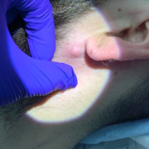
How Suspicious Moles Are Assessed in Clinic
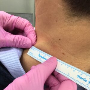
Assessment of a suspicious mole is a structured clinical process rather than a single visual judgement. At City Dermatology Clinic, initial evaluation is dermatologist-led, with surgical input considered where appropriate.
The assessment typically begins with a detailed clinical review. This includes discussion of when the mole first appeared, whether it has changed over time, and whether any symptoms such as bleeding, itching, or discomfort have developed. Personal and family history of skin cancer, sun exposure patterns, and previous mole removals may also be relevant to the overall assessment.
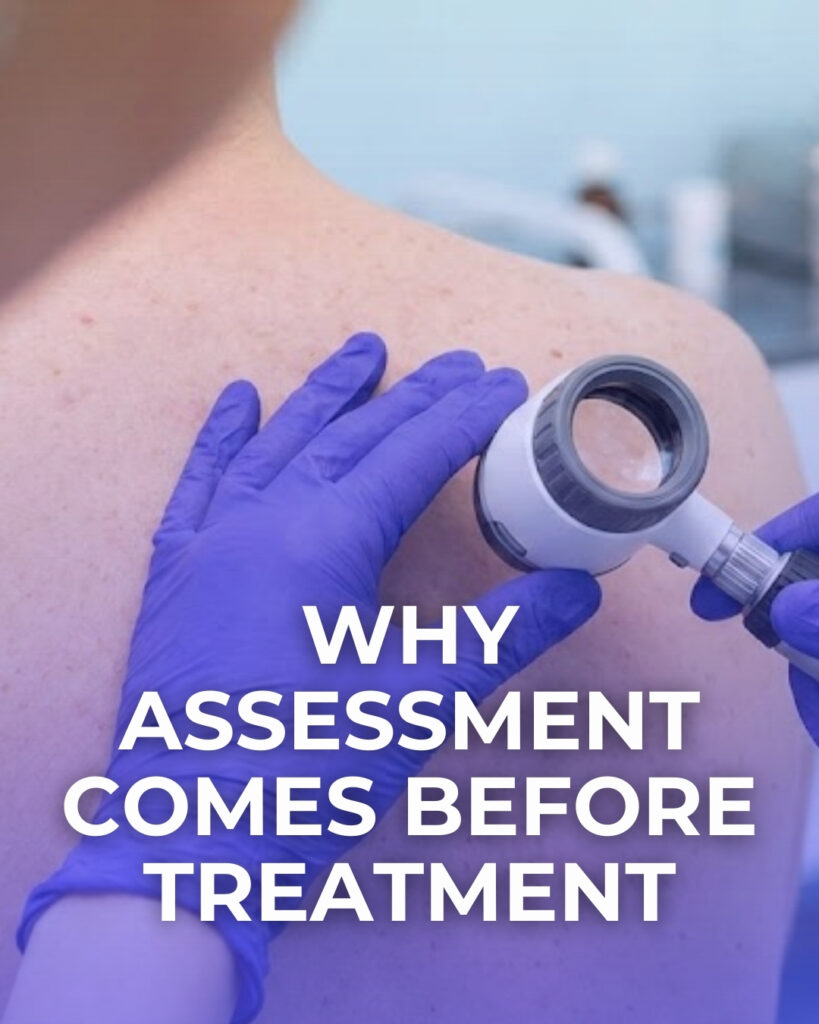
A physical examination follows, using dermoscopy. Dermoscopy allows dermatologists to examine pigment networks and subsurface structures that are not visible to the naked eye. These features can help distinguish between lesions that appear similar on the surface but behave differently at a cellular level. Dermoscopic findings are interpreted alongside clinical history rather than in isolation.
At this stage, several outcomes are possible:
- Reassurance, where the mole shows benign features
- Monitoring, if changes are subtle and removal is not immediately indicated
- Removal, where diagnostic clarification or symptom management is required
Importantly, removal is not positioned as the default outcome. The purpose of assessment is to decide whether intervention is appropriate and, if so, to determine the safest and most suitable approach for that individual mole and patient.
The Role of Dermatologists and Plastic Surgeons in Mole Assessment and Removal
Management of suspicious moles often benefits from input across more than one specialty. Dermatologists and plastic surgeons bring different but complementary expertise to assessment and treatment decisions.
Dermatologists are central to diagnosis and triage. At City Dermatology Clinic, mole assessments are performed by consultant dermatologists such as Dr Andreea Anton, whose practice includes mole checks, dermoscopic assessment, and skin cancer screening. Dermatologists focus on identifying concerning features, deciding whether histological analysis is required, and advising on appropriate next steps.
Plastic surgeons are involved when surgical planning, anatomical complexity, or cosmetic considerations are relevant. Lesions on the face, neck, or other visible areas may require a more tailored surgical approach to balance diagnostic needs with healing and scarring considerations. Consultant plastic surgeons including Mr Samim Ghorbanian, Mr Georgios Pafitanis, and Dr Jana Torres‑Grau routinely manage skin lesion excisions as part of their broader reconstructive and skin cancer–related practice.
This collaborative model allows decisions to be based on:
- Diagnostic clarity
- Lesion location and size
- Surgical complexity
- Anticipated healing and scarring
Where removal is advised, the technique and closure method are selected according to these factors, rather than a one-size-fits-all approach. Histology is arranged when clinically indicated, and results are reviewed as part of the ongoing care pathway.
By combining dermatologist-led assessment with access to experienced plastic surgeons, the clinic provides a structured, specialist approach to suspicious mole management that prioritises accuracy, safety, and appropriate surgical planning.
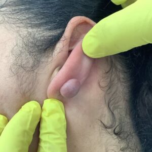
Removal Options for Suspicious Moles
When removal of a suspicious mole is advised, the approach is selected based on clinical need rather than convenience or routine preference. The primary objective is diagnostic accuracy, with cosmetic and functional considerations addressed alongside this.
The most commonly used removal methods include:
Excision Biopsy
This involves removing the mole in full, including a margin of surrounding skin where appropriate. Excision is often used when diagnostic clarity is required or when the mole’s features warrant complete removal for histological examination. The wound is typically closed with sutures, and the method of closure is chosen based on the location of the lesion and surrounding skin tension.
Shave Removal or Shave Biopsy
In selected cases, a superficial shave technique may be appropriate. This is usually considered for lesions that appear confined to the upper layers of the skin and where full-thickness excision is not required for diagnostic purposes. The decision to use a shave technique is made cautiously and only after assessment.
Technique Selection
The choice of removal method depends on several factors, including:
- Dermoscopic findings
- Lesion depth and location
- Diagnostic requirements
- Healing and scarring considerations
No single technique is suitable for all moles. Part of specialist assessment is determining whether removal is needed at all, and if so, how it should be performed to balance diagnostic accuracy with safe healing.
Histology and What Happens After Removal
When a suspicious mole is removed, the tissue is often sent for histological analysis. Histology involves examining the removed tissue under a microscope to assess cellular features and confirm the nature of the lesion.
Not every removed mole requires histology, but it is commonly recommended when:
- There is diagnostic uncertainty
- The mole has changed over time
- Clinical or dermoscopic features warrant confirmation
Histology results typically fall into one of several broad categories, which are discussed with the patient once available. The role of the clinician at this stage is to explain the findings clearly and outline any recommended next steps, if applicable.
Follow-up after removal may include:
- Review of wound healing
- Discussion of histology results
- Further management or monitoring, depending on findings
Importantly, histology is used to inform care rather than to assume outcomes. Many suspicious moles are ultimately confirmed as benign, and the process exists to ensure that uncertainty is managed safely and appropriately.
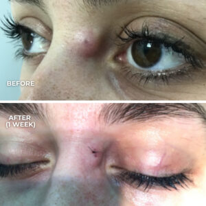
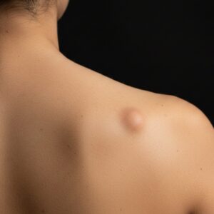
Recovery, Scarring, and Healing Expectations
Recovery following mole removal varies between individuals and depends on several factors, including the size and location of the lesion, the removal technique used, and individual healing characteristics.
After removal, it is normal for the area to go through a healing phase that may include mild swelling, redness, or temporary discomfort. Where sutures are used, these are typically removed or dissolve according to the anatomical site and closure method. Wound care instructions are provided to support healing and reduce the risk of complications.
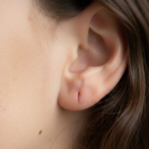
Scarring is always a possibility when the skin is broken. The eventual appearance of a scar depends on:
- The location of the mole
- The depth of removal
- Skin type and healing response
- Tension across the wound
Moles removed from areas such as the face, neck, or upper chest may require more careful surgical planning due to visibility and skin movement. This is one reason why surgical input can be relevant in selected cases. Expectations around healing and scarring are discussed during consultation so that decisions are informed and realistic.
Who May Benefit from a Specialist Mole Assessment
A specialist assessment may be appropriate for individuals who notice changes in an existing mole, develop a new pigmented lesion, or feel uncertain about the nature of a mole.
Common reasons patients seek assessment include:
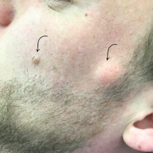
- A mole that has changed in size, shape, or colour
- New symptoms such as bleeding, itching, or crusting
- A mole that looks different from others on the body
- Ongoing uncertainty or concern
It is also common for patients to request assessment following advice from a GP or after difficulty obtaining timely specialist review elsewhere. In many cases, assessment provides reassurance rather than a recommendation for removal.
Seeking assessment does not commit a patient to treatment. The purpose of consultation is to clarify whether any action is needed and, if so, to explain appropriate options.
Booking a Specialist Assessment in London
Assessment of suspicious moles is based on clinical evaluation rather than assumption. A consultation allows a specialist to examine the mole, discuss any relevant history, and explain whether monitoring, removal, or no further action is appropriate.
At City Dermatology Clinic, assessments are led by consultant dermatologists, with access to plastic surgeons when surgical planning or anatomical considerations are relevant. This allows care decisions to be tailored to the individual lesion and patient.
If you have noticed changes in a mole or would like professional reassurance, booking a consultation provides an opportunity to have the area assessed and to discuss appropriate next steps based on clinical findings.